Article Contents
 Return home
From Clinical Observation to Neurophysiologic Hypothesis: Twenty-Five Years of Retinal Cholinergic Modulation in Stargardt Disease
Gerard M. Nolan, MD, FACS September 2026
Abstract
A patient with Stargardt disease experienced visual improvement following topical Phospholine Iodide® / Echothiophate Iodide (PI/ECHO) treatment between 2001 and 2006, with improvement independently documented by two ophthalmologists in another state. Recent genetic testing subsequently confirmed the diagnosis of Stargardt disease.
Stargardt disease is an inherited retinal degeneration characterized predominantly by progressive dysfunction and loss of photoreceptors and retinal pigment epithelium, with preservation of portions of the inner retinal circuitry. In 2001, an unexpected visual response was observed in a 27-year-old woman with advanced Stargardt disease and legal blindness following treatment with dilute topical PI/ECHO, a long-acting acetylcholinesterase inhibitor (AChEI). The initial therapeutic rationale included the possibility that pharmacologically induced miosis might improve vision through an optical pinhole effect. Subsequent observations, however, were not readily explained by pupil constriction alone.
The patient’s pretreatment visual dysfunction had been independently documented in 1997. Color discrimination was markedly abnormal, dark-adaptation thresholds were 18 dB OD and 22 dB OS, and visual acuity had reached the level of legal blindness. Following initiation of dilute PI/ECHO, she reported improvement in color vision, night vision, reading, mobility, and general visual function. Independent reevaluation in 2003 noted improvement in both the treated and untreated eyes. In 2004, repeat testing demonstrated completely normal Farnsworth D-15 color responses and improvement of dark-adaptation thresholds to 6 dB OD and 5 dB OS. In contrast, objective electrophysiologic abnormalities attributable to Stargardt disease persisted.
These observations initiated a 25-year clinical investigation into whether augmentation of cholinergic signaling might influence residual retinal or central visual processing. Subsequent observations, including bilateral improvement following unilateral treatment, persistence of acuity improvement after pharmacologic reversal of miosis, and visual improvement in an aniridic eye, further challenged a purely optical explanation.
This historical clinical experience does not establish efficacy or prove a neurophysiologic mechanism. Rather, the dissociation between visual function, pupillary effects, and electrophysiologic or structural retinal disease raises a testable hypothesis: enhancement of cholinergic signaling may modify information processing within surviving retinal and/or central visual pathways despite persistent outer-retinal pathology.
Introduction
Stargardt disease is the most common inherited juvenile macular dystrophy and is most frequently associated with pathogenic variants in ABCA4. Accumulation of toxic retinoid byproducts, retinal pigment epithelial dysfunction and progressive photoreceptor degeneration result in central visual loss, impaired color discrimination, and, in more advanced disease, abnormalities of dark adaptation and broader retinal function.
Although the primary pathology involves the outer retina and retinal pigment epithelium, substantial elements of the inner retinal neural network may remain anatomically present. This raises an important neurophysiologic question:
Can surviving retinal circuitry be pharmacologically modulated to improve the processing of degraded photoreceptor input without reversing the underlying retinal degeneration?
An unexpected clinical observation approximately 25 years ago led to investigation of this possibility.
PI/ECHO is a long-acting acetylcholinesterase inhibitor. By inhibiting acetylcholinesterase, it increases the availability and duration of endogenous acetylcholine. Applied topically to the eye, it produces prominent miosis, initially suggesting a straightforward optical explanation for any improvement in acuity. Over subsequent years, however, several observations emerged that were increasingly difficult to reconcile with a pinhole mechanism alone.
This article describes the clinical observations that led from an initial optical hypothesis to a broader hypothesis involving cholinergic modulation of retinal and potentially central visual processing.
The Pretreatment Clinical Record: 1997
The index patient (A260) was a young woman who developed progressive loss of central acuity, color discrimination, and night vision. By early 1997, she described substantial difficulty reading because portions of words appeared to be missing, marked reduction in color vision, photophobia, and progressive impairment of night vision.
On January 29, 1997, Jonathan S. Lyons, MD, performed an independent ophthalmologic and electrodiagnostic evaluation.1 Distance visual acuity was approximately finger counting at 5 feet in each eye, and near visual acuity was 20/200 OU. Farnsworth D-15 testing demonstrated markedly diminished color discrimination. Funduscopic examination demonstrated bull’s-eye pigmentary changes involving the central retinal pigment epithelium with surrounding retinal pigment epithelial abnormalities. Full-field electroretinography was essentially normal, consistent with retinal disease predominantly involving the macular region. Dark-adaptation testing, however, was markedly abnormal. Thresholds were 18 dB OD and 22 dB OS, compared with a reported normal value of less than 5 dB.
On February 6, 1997, Daniel Finkelstein, MD, at the Wilmer Ophthalmological Institute of the Johns Hopkins University School of Medicine independently evaluated the patient. Her fundus examination and fluorescein angiographic findings were considered compatible with fundus flavimaculatus/Stargardt disease. Her visual acuity was considered to have reached the level of legal blindness in each eye.2 The diagnosis of Stargardt disease was established clinically in 1997, before molecular genetic testing became routinely incorporated into the evaluation of inherited retinal disease. Genetic testing was not performed at that time. The patient and her father were advised that there was no available treatment for Stargardt disease, and low-vision rehabilitation and additional baseline evaluations were recommended.
On August 29, 2026, contemporary inherited retinal disease genetic testing, including analysis of ABCA4, was submitted to provide molecular characterization of the longstanding clinical diagnosis. Subsequent testing identified two pathogenic ABCA4 variants and one pathogenic low-penetrance ABCA4 variant, providing molecular support for the clinical diagnosis of ABCA4-associated Stargardt disease; variant phase has not yet been established (see Appendix B).
These records are important because they provide independent documentation of substantial visual dysfunction several years before initiation of the treatment described below.
Original Clinical Observation: 2001
On June 18, 2001, at age 27, the patient had advanced Stargardt disease with severe central visual impairment affecting reading, color perception, navigation, and independence. Dilute topical PI/ECHO was administered off-label. A concentration of approximately 0.015% was used immediately before overnight sleep. See Appendix A.
The initial rationale centered on pharmacologically induced miosis. Reduction in pupil diameter can increase depth of focus and decrease optical aberrations, producing a pinhole-like improvement in selected patients. It was therefore reasonable initially to attribute any visual change to an optical mechanism. The magnitude and character of the patient’s response, however, were unexpected. She reported improvement not only in distance and near acuity but also in color perception, night vision, reading ability, mobility, and daily visual function. These changes permitted substantially greater functional independence. Continued treatment was associated with maintenance of the visual response. Interruption of therapy was subsequently associated with loss of improvement, while reinstitution of treatment was followed by return of visual benefit.5
The observation was described in a manuscript prepared in 2003 and was also discussed publicly by the Foundation Fighting Blindness that year.6 Even at that early stage, two possible explanations were considered:
- An optical pinhole effect produced by miosis
- A retinal mechanism in which increased acetylcholine might amplify or enhance residual visual signaling
Independent Reevaluation: 2003
In March 2003, the patient returned to Daniel Finkelstein, MD, at Wilmer for independent reevaluation.3 Of particular importance, Finkelstein acknowledged the improvement that had occurred during treatment and noted that both the treated and untreated eye had improved.4 This observation became important to the subsequent mechanistic interpretation.
If improvement resulted exclusively from miosis in the treated eye, comparable improvement in an untreated fellow eye would be difficult to explain through a local optical pinhole mechanism alone. A bilateral response raised the possibility of systemic, consensual neural, retinal or central effects.
Finkelstein also expressed interest in evaluating additional patients before and after treatment with objective testing capable of documenting changes in addition to subjective visual responses.4 The 2003 evaluation therefore represents an important transition in the history of these observations: the clinical question was no longer simply whether the patient perceived improvement, but whether the pattern of improvement could be reconciled with the presumed optical mechanism.
Seven-Year Comparative Evaluation: 2004
On February 18, 2004, Lyons reevaluated the patient approximately seven years after his original examination. The patient reported that during the preceding two years, concurrent with treatment using dilute PI/ECHO, her color vision and night vision had improved.8 Visual acuity measured 20/200 OU. Slit-lamp examination was normal, while funduscopic examination continued to demonstrate bilateral atrophic macular changes.
The most striking difference involved color discrimination. Farnsworth D-15 testing now demonstrated completely normal responses, compared with the significant color defect documented in 1997. Dark adaptation also showed a substantial change.
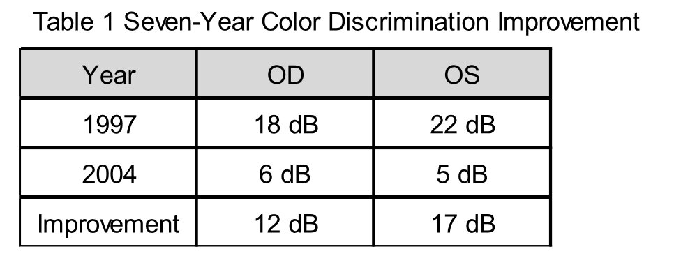
Thus, dark-adaptation thresholds approached the approximately 3 dB value reported as normal for her age at the 2004 examination. Importantly, electrophysiologic evidence of Stargardt disease persisted. Multifocal electroretinography demonstrated severe reduction in waveform amplitude throughout much of the central 30-degree field in both eyes, consistent with Stargardt disease. Full-field electroretinalgram (ERG) amplitudes and implicit times remained normal under photopic and scotopic conditions and were similar to the patient’s previous testing.
Lyons concluded that the follow-up evaluation demonstrated improvement in color vision and dark adaptation compared with the previous examination, while objective electrophysiologic testing remained unchanged.8 This distinction is critical. The findings do not demonstrate restoration of diseased photoreceptors or reversal of the retinal degeneration. Instead, they demonstrate improved performance on functional visual testing despite persistent evidence of the underlying retinal disorder.
From an Optical to a Neurophysiologic Hypothesis
Miosis remained the most obvious alternative explanation for improved visual acuity. A smaller pupil can reduce optical aberrations and increase depth of focus. The original hypothesis therefore appropriately included a pinhole mechanism.
Over time, however, several clinical observations became increasingly difficult to explain by pupil size alone:
- Improvement occurred in an untreated fellow eye following unilateral treatment
- Pharmacologic reversal of miosis with mydriatic agents did not immediately abolish the visual acuity improvement in subsequent observations
- The index patient substantial changes in color discrimination and dark adaptation, visual functions not readily accounted for by a simple pinhole effect
- Visual improvement was subsequently observed in an aniridic patient, in whom an iris-mediated pinhole mechanism could not account for the response
Taken together, these observations suggested that miosis might accompany treatment without necessarily being the sole mediator of visual improvement.
Retinal Cholinergic Modulation
Acetylcholine is an important neurotransmitter within the retina. Cholinergic starburst amacrine cells participate in complex signaling networks involving bipolar, amacrine and retinal ganglion cells. These networks contribute to temporal and spatial processing and to the transformation of photoreceptor-derived signals into the retinal ganglion-cell output transmitted to the brain.
In Stargardt disease, severe photoreceptor and retinal pigment epithelial abnormalities can coexist with surviving inner-retinal neurons. The persistence of these neural elements raises the possibility that pharmacologic modulation could alter how residual photoreceptor signals are processed.
PI/ECHO irreversibly inhibits acetylcholinesterase, prolonging the action of acetylcholine. The working hypothesis arising from these clinical observations is that enhanced cholinergic activity may modify signaling within surviving retinal circuitry and potentially influence downstream central visual processing. Under this model, treatment would not regenerate lost photoreceptors or restore atrophic retinal pigment epithelium. Rather, surviving neural networks might process limited or degraded retinal input more effectively. This interpretation could explain an apparent paradox in the index patient: substantial functional improvement in color discrimination and dark adaptation despite persistent macular abnormalities and unchanged electrophysiologic evidence of retinal disease.
It remains a hypothesis rather than a demonstrated mechanism.
Twenty-Five Years of Clinical Observation
The original patient observation led to a broader clinical investigation of dilute topical PI/ECHO in patients with Stargardt disease. Subsequent clinical experience included larger retrospective cohorts and observations involving genetically confirmed ABCA4 disease. Across these experiences, improvement was observed in distance acuity, near acuity and color discrimination in a proportion of treated patients.6
The accumulated observations also produced several recurring mechanistic findings:
- Improvement could occur bilaterally following unilateral administration;
- Visual benefit could persist despite reversal of miosis;
- Discontinuation could be associated with loss of benefit
- Retreatment with renewed improvement;
- Improvement could occur in the absence of an iris.
These findings do not substitute for randomized, masked, controlled clinical trials. The experience spans different periods of clinical practice, testing methods, disease severity, and study designs. Placebo effects, learning effects, regression to the mean, optical factors, and other sources of bias cannot be completely excluded. The historical value of the observations lies instead in the consistency of an unexpected clinical phenomenon and in the generation of a biologically testable hypothesis.
Discussion
The 1997 – 2004 records provide an unusual longitudinal window into the evolution of the original observation. Before treatment, two independent ophthalmologists documented a clinical phenotype consistent with advanced Stargardt disease and severe visual dysfunction. Molecular genetic testing was not performed at that time.
Color discrimination and dark adaptation were markedly impaired, and the patient’s visual acuity met criteria for legal blindness. During treatment, color discrimination normalized on Farnsworth D-15 testing and dark-adaptation thresholds changed from 18 and 22 dB to 6 and 5 dB. These findings were documented by the same independent electrophysiologic examiner who had performed the pretreatment evaluation seven years earlier. At the same time, electrophysiologic abnormalities did not demonstrate corresponding recovery. Rather than weakening the observation, this dissociation may help define it. The clinical phenomenon appears to concern visual function rather than anatomical retinal restoration.
A neurophysiologic interpretation would predict precisely such a possibility: altered processing of residual information without regeneration of the damaged outer retina.
The bilateral response after unilateral treatment similarly raises questions about the site of action. A local ocular pharmacologic effect remains possible, but the observation also invites investigation of systemic absorption, interocular neural interactions, retinal network modulation and central visual processing.
Modern neuroscience provides tools that were not readily available when the original observations were made. Adaptive-optics retinal imaging, quantitative multifocal ERG, pattern and multifocal visual evoked potentials, pupillometry, microperimetry, quantitative color and contrast testing and functional magnetic resonance imaging could now be combined to determine whether changes occur at the photoreceptor, inner-retinal, optic pathway or visual cortical level.
Prospective studies should specifically separate optical from neural effects. Pupil diameter should be measured continuously, acuity should be tested under controlled artificial apertures, and pharmacologic reversal of miosis should be incorporated when appropriate. Unilateral administration with masked bilateral testing could examine interocular effects. ERG and visual evoked potentials could help distinguish retinal from post-retinal responses.
The central question generated by this 25-year clinical experience is therefore broader than whether PI/ECHO improves visual acuity in Stargardt disease. It is:
Can cholinergic modulation enhance residual visual processing in a degenerating human retinal system?
Limitations
These observations must be interpreted cautiously. The index patient’s original 1997 diagnosis was based on clinical, funduscopic, fluorescein angiographic, and electrophysiologic findings rather than molecular genetic confirmation. In August 2026, comprehensive inherited retinal disease genetic testing provided molecular support for the longstanding clinical diagnosis of ABCA4-associated Stargardt disease.
The original experience began with a single patient and was not derived from a randomized controlled trial. Some outcome measures, including color and dark-adaptation testing, have psychophysical components and therefore depend on patient responses. The 2004 electrophysiologic examination did not demonstrate objective improvement corresponding to the functional changes.
The bilateral response following unilateral treatment is mechanistically provocative but does not establish a central nervous system mechanism. Systemic absorption or other pharmacologic explanations require consideration. Similarly, persistence of visual improvement following reversal of miosis and improvement in an aniridic patient argue against a purely pupillary explanation but do not, by themselves, establish the anatomical site or molecular mechanism responsible for the response. Accordingly, the accumulated findings should be viewed as hypothesis-generating clinical observations requiring prospective experimental validation.
Conclusions
A clinical observation beginning approximately 25 years ago suggested that dilute topical PI/ECHO might be associated with improved visual function in a patient with advanced Stargardt disease.
Independent records are particularly informative. Before treatment, the patient demonstrated legal blindness, markedly abnormal color discrimination, and dark-adaptation thresholds of 18 dB OD and 22 dB OS. During treatment, color discrimination became normal on Farnsworth D-15 testing and dark-adaptation thresholds improved to 6 dB OD and 5 dB OS. These functional changes occurred despite persistent macular pathology and without corresponding improvement in electrophysiologic testing.
Subsequent observations – including bilateral improvement following unilateral treatment, persistence of visual benefit despite pharmacologic reversal of miosis and improvement in an aniridic eye – further challenged the original assumption that the response could be explained entirely by a pinhole effect.
The resulting hypothesis is not that cholinergic modulation regenerates the degenerating retina. Rather, augmentation of acetylcholine signaling may alter the processing of residual visual information within surviving retinal and/or central neural pathways.
After 25 years, the significance of the original observation may therefore extend beyond Stargardt disease. It raises a broader neuroscience question:
Can pharmacologic neuromodulation improve function in a partially damaged sensory system by enhancing the performance of neural circuitry that remains?
That question is now experimentally testable and warrants rigorous prospective investigation.
Footnoted Mechanistic Paragraph
The historical observations from 1997 through 2004 preceded much of the contemporary molecular and neurophysiologic understanding that now provides a framework in which they can be reconsidered. The patient’s Stargardt phenotype and color-vision dysfunction were independently documented before treatment,1,2 followed by functional improvement after initiation of dilute topical PI/ECHO in 2001.3,4 The response was described in contemporaneous correspondence,3 in a manuscript submitted in 20035 and publicly by the Foundation Fighting Blindness that same year.7 Most importantly, independent electrophysiologic re-evaluation in 2004 documented normalization of Farnsworth D-15 color testing compared with the abnormal response recorded seven years earlier.8
Modern work has established that Stargardt disease primarily affects photoreceptor/RPE physiology through abnormal ABCA4-mediated retinoid handling and accumulation of toxic bisretinoids.9-12 At the same time, the retina is a complex neural network in which acetylcholine released principally by starburst amacrine cells can modulate bipolar, amacrine, and ganglion-cell signaling.13-15 These findings provide a biologically plausible basis for the hypothesis that acetylcholinesterase inhibition could influence residual retinal signal processing independently of an optical pinhole effect. They do not, by themselves, establish PI/ECHO as a treatment for Stargardt disease; rather, they provide a mechanistic framework within which the longitudinal clinical observations can be examined.
Appendix A
Historical Index Patient: Longitudinal Visual Response to Topical Echothiophate Iodide
Background
The index patient (A260) was a woman diagnosed with Stargardt disease at age 19 after initially experiencing difficulty distinguishing colors at approximately age 17, followed by progressive central and night-vision loss. The diagnosis was subsequently confirmed by retinal specialists, including evaluation at The Johns Hopkins Wilmer Eye Institute. This patient provided the initial clinical observation that led to the subsequent investigation of topical cholinergic modulation of visual function in Stargardt disease.
Baseline testing before treatment included distance best corrected visual-acuity (BCVA), near BCVA, color vision, pupillary examination, and fluorescein angiography. The patient demonstrated severe central visual impairment with eccentric fixation. Fluorescein angiography demonstrated retinal pigment epithelial atrophy localized predominantly to the macula, subsequently classified as Stage III disease.
Treatment with dilute topical PI/ECHO (0.015%) was initiated on June 18, 2001. The right eye was treated initially, followed by treatment of the left eye and subsequently alternating-eye administration. The original September 2003 manuscript documented the early treatment protocol and clinical response.
Longitudinal Visual Function
Initial Treatment Response
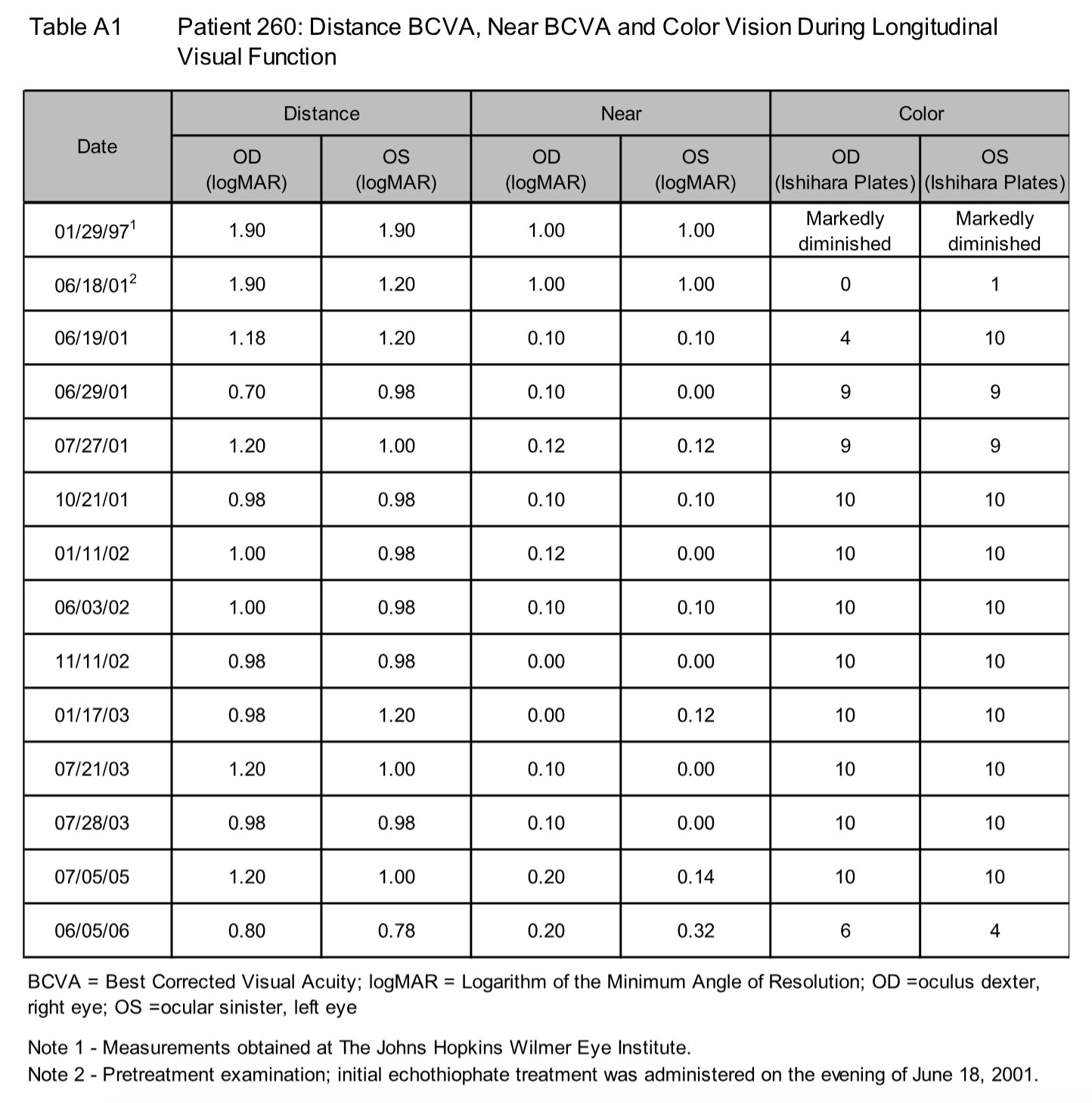
The pretreatment examination on June 18, 2001 demonstrated distance BCVA of 1.90 logMAR OD and 1.20 logMAR OS, near BCVA of 1.00 logMAR in each eye, and color discrimination of 0 Ishihara plates OD and 1 plate OS. Approximately 24 hours after the first treatment of the right eye, distance BCVA in that eye improved from 1.90 to 1.18 logMAR. Near BCVA improved bilaterally from 1.00 to 0.10 logMAR. Color discrimination increased from 0 to 4 plates in the treated right eye and, notably, from 1 to 10 plates in the untreated left eye.
The original 2003 report similarly described rapid improvement in distance and near vision and marked improvement in color discrimination following treatment.
Long-Term Course
Serial examinations demonstrated persistence of improved visual function over subsequent years. Color discrimination reached 10/10 Ishihara plates bilaterally during much of the follow-up period. Distance and near acuity fluctuated but generally remained improved relative to the severe pretreatment measurements.
At the July 5, 2005 examination, distance BCVA was 1.20 logMAR OD and 1.00 logMAR OS, near BCVA was 0.20 logMAR OD and 0.14 logMAR OS, and color vision remained 10/10 plates in each eye. At the final examination included in this appendix, June 5, 2006, distance BCVA measured 0.80 logMAR OD and 0.78 logMAR OS, while near BCVA measured 0.20 logMAR OD and 0.32 logMAR OS. Color discrimination at that examination was 6/10 plates OD and 4/10 plates OS. Thus, the longitudinal response was not uniform across all visual measures, particularly color discrimination at the final examination.
Treatment Interruption and Rechallenge
During the approximately five-year treatment period, the patient experienced an inadvertent two-week interruption of PI/ECHO therapy after failing to bring the medication on her honeymoon. According to the contemporaneous clinical history, after approximately five days without treatment, visual function declined to the patient’s previous level of legal blindness and color vision was lost. Following resumption of PI/ECHO, visual acuity and color discrimination improved and again stabilized.
This uncontrolled withdrawal-and-rechallenge observation does not establish causality but is noteworthy because deterioration occurred following treatment withdrawal and improvement was reported following re-exposure to the medication.
Historical and Mechanistic Significance
This index case is important to the 25-year clinical history because the response was documented across multiple domains of visual function – distance acuity, near acuity, color discrimination and functional vision – and was followed longitudinally for approximately five years. The early response was documented contemporaneously in the 2003 manuscript rather than reconstructed retrospectively.
Of particular interest was the bilateral response after initial unilateral treatment, including improvement in the untreated eye, together with the marked changes in color discrimination. These observations were difficult to attribute solely to an optical pinhole effect from miosis and contributed to the subsequent hypothesis that acetylcholinesterase inhibition could influence retinal and/or post-retinal visual processing.
The findings from this single patient should be considered historical and hypothesis-generating rather than proof of therapeutic efficacy. Nevertheless, the extended longitudinal record documents the clinical observation from which the subsequent investigation of retinal cholinergic modulation in Stargardt disease developed.
Appendix B
Genetic Confirmation
In August 2026, comprehensive inherited retinal disease genetic testing identified two pathogenic ABCA4 variants, c.1622T>C (p.Leu541Pro) and c.3113C>T (p.Ala1038Val), together with a pathogenic low-penetrance ABCA4 variant, c.5882G>A (p.Gly1961Glu). These findings provide molecular support for the longstanding clinical diagnosis of ABCA4-associated Stargardt disease. The phase of the variants has not yet been established; therefore, it remains undetermined whether causative variants are present on opposite chromosomes.
References
- Lyons JS. Electrodiagnostic consultation. Silver Spring, MD: Jonathan S. Lyons, MD, Ophthalmology & Ophthalmic Surgery; January 29, 1997. Unpublished clinical report.
- Finkelstein D. Letter to Mary S. Carlson, MD. The Wilmer Ophthalmological Institute, The Johns Hopkins University School of Medicine, Baltimore, MD; February 6, 1997. Unpublished clinical correspondence.
- Finkelstein D. Letter to Gerard Nolan, MD. The Wilmer Ophthalmological Institute, The Johns Hopkins University School of Medicine, Baltimore, MD; March 14, 2003. Unpublished correspondence.
- Finkelstein D. Letter to Gerard Nolan, MD. The Wilmer Ophthalmological Institute, The Johns Hopkins University School of Medicine, Baltimore, MD; March 25, 2003. Unpublished clinical correspondence.
- Nolan GM. The successful management of Stargardt’s disease using topical diluteed echothiophate iodide. Manuscript submitted for publication; September 2003, www.stargardtdiseasetreatment.com.
- Nolan, GM,Cholinergic Maculation in Hereditary Retnal Disease: Stargardt Disease, Manuscript submitted for publication, June, 2026, www.stargardtdiseasetreatment.com.
- Foundation Fighting Blindness. A Glaucoma Drug for Stargardt Disease: The Use of Dilute PI/ECHOthiophate Iodide Eyedrops for Stargardt Disease. Stargardt Disease Research. 2003. Archived web publication.
- Lyons JS. Electrodiagnostic report. Silver Spring, MD: Jonathan S. Lyons, MD, LLC; February 18, 2004. Unpublished clinical report.
- Molday RS, Zhong M, Quazi F. The role of the photoreceptor ABC transporter ABCA4 in lipid transport and Stargardt macular degeneration. Biochim Biophys Acta. 2009;1791:573-583.
- Quazi F, Lenevich S, Molday RS. ABCA4 is an N-retinylidene-phosphatidylethanolamine and phosphatidylethanolamine importer. Nat Commun. 2012;3:925.
- Molday RS, Garces FA, Scortecci JF, Molday LL. Structure and function of ABCA4 and its role in the visual cycle and Stargardt macular degeneration. Prog Retin Eye Res. 2022;89:101036. doi:10.1016/j.preteyeres.2021.101036.
- Lenis TL, Hu J, Ng SY, et al. Expression of ABCA4 in the retinal pigment epithelium and its implications for Stargardt macular degeneration. Proc Natl Acad Sci U S A. 2018;115. doi:10.1073/pnas.1802519115.
- Baldridge WH. Optical recordings of the effects of cholinergic ligands on neurons in the ganglion cell layer of mammalian retina. J Neurosci. 1996;16:5060-5072.
- Yamada ES, Dmitrieva N, Keyser KT, Lindstrom JM, Hersh LB, Marshak DW. Synaptic connections of starburst amacrine cells and localization of acetylcholine receptors in primate retinas. J Comp Neurol. 2003;461:76-90.
- O’Malley DM, Masland RH. Co-release of acetylcholine and gamma-aminobutyric acid by a retinal neuron. Proc Natl Acad Sci U S A. 1989;86:3414-3418.
Abbreviations and Acronyms:
AChEI = Acetylcholinesterase Inhibitor; BCVA = Best Corrected Visual Acuity; ERG = Electroretinalgram; logMAR = Logarithm of the Minimum Angle of Resolution; OD = oculus dexter, right eye; OS = ocular sinister, left eye; OU = Oculus Uterque, both eyes; PI/ECHO = Phospholine Iodide® / Echothiophate Iodide